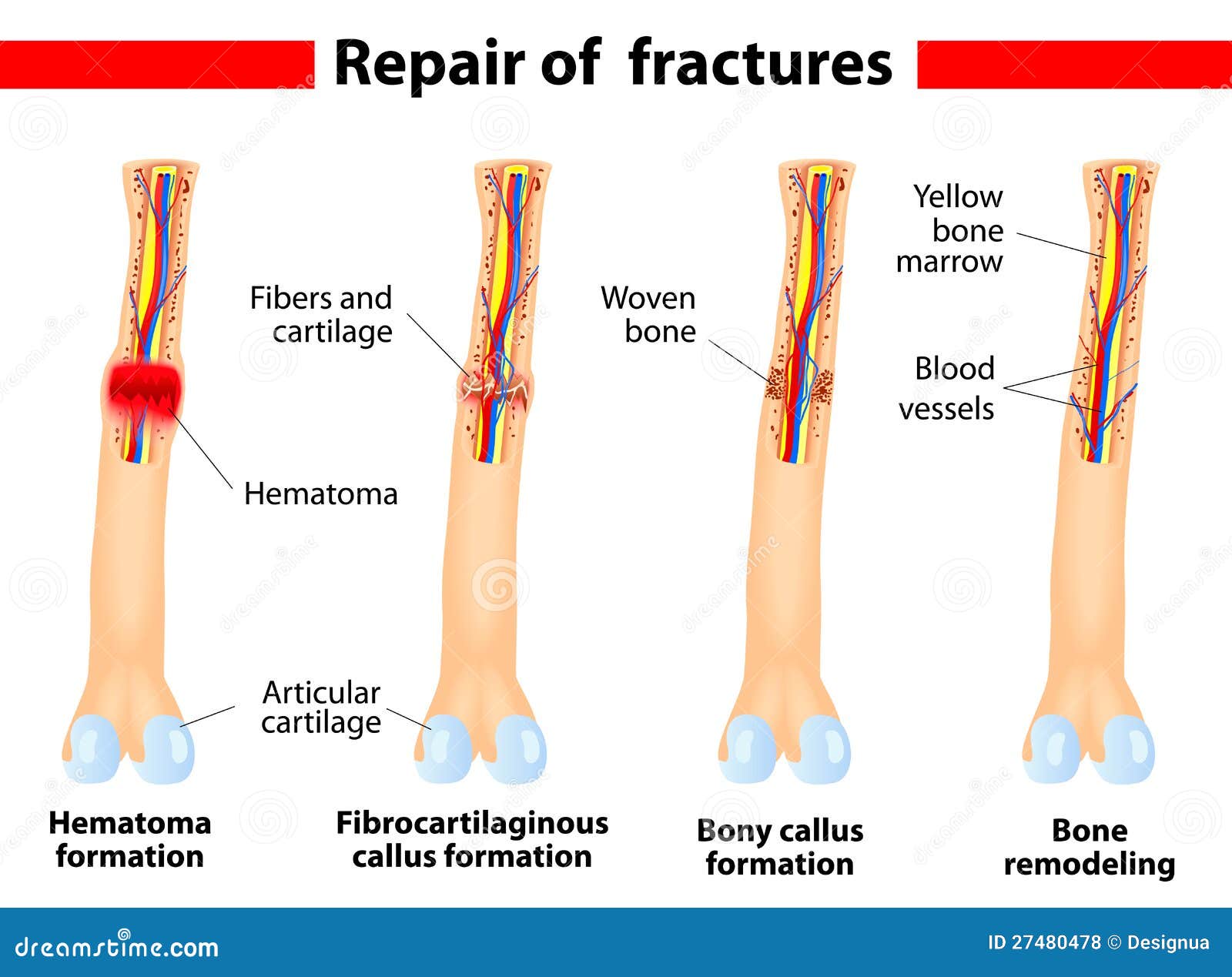
fracture healing process

[music playing] narrator: some have describedthe skeletal system as the infrastructure of the humanbody, providing a rigid framework that offers protectionand support, as well as attachment sites for thetendons and muscles that are essential for locomotion. the skeletal system is comprisedof 206 bones, each of which is formed through aprocess known as ossification. bone development and remodelingtakes place on a
continual basis, from theprenatal period and early childhood through adulthood, aswell as during the healing process that occurs followingbone fractures. ossification can take one oftwo forms, depending on the type of bone that is forming. in the case of flat bones,like those of the ribs, scapula, pelvis, and skull, theossification process is known as intramembranousossification. the process is initiated bymesenchymal stem cells, which
are cells that have yet tobe differentiated into a particular cell type. eventually, centrally locatedmesenchymal cells within the embryonic fibrous connectivetissue differentiate into bone-forming cellsor osteoblasts. this process constitutes theformation of what are called ossification centers. in the matrix formation phasethat follows, the osteoblasts begin to lay down osteoid, whichis the organic part of
the bone, consistingof collagen fibers. some of the osteoblasts areentrapped within the osteoid and are then said tobe osteocytes. the osteoid then calcifies andforms slender needle-like structures of spongy bonecalled spicules, which aggregate in the form of smallsupporting beams or trebeculae. meanwhile, the blood vessels onthe outside of the spongy bone condense and form theperiosteum, the fibrous sheet
that covers bone. as the trebeculae thicken, aninterconnected network forms, resulting in what's calledwoven bone, which is characterized by a somewhathaphazard organization of collagen fibers and ismechanically weak. eventually, this leads to theformation of lamellar bone, around a newly formedspongy bone. this lamellar or compact bonehas regular, parallel alignment of collagen in theform of sheets or lamellae and
is mechanically quite strong. while flat bones are formedthrough intramembraneous ossification, most bones inthe body, such as the long bones of the legs and arms,are formed in a somewhat different manner known asendochondral ossification. this type of bone developmentinvolves cartilage models, which are eventually replacedwith bony tissue. the process begins in thedeveloping fetus and can be divided into several stages.
around the eighth week ofdevelopment, chondroblasts begin secreting a cartilaginousmatrix that will form the hyaline cartilage modelfor bone development. as with other hyalinecartilage tissues, chondrocytes are trapped inlacunae and a perichondrium surrounds the model. the next stage begins whenchondrocytes within the center of the model hypertrophy andbegin to resorb some of the surrounding cartilage matrix.
as these chondrocytes enlarge,the matrix begins to calcify and chondrocytes begin to die,as nutrients cannot diffuse through the newly-formedcalcified matrix. next, stem cells within theperichondrium divide to form osteoblasts and a compact bonecollar is formed around the calcified cartilage shaft. a periosteal bud consisting ofcapillaries and osteoblasts invades the core of thecartilaginous shaft, forming a primary ossification center.
the remains of the calcifiedcartilage serve as a template on which osteoblastscan build bone. bone development extends towardsthe epiphyses from the most primary ossificationcenters are formed by the 12th week of development. the same basic process ofprimary ossification is repeated in the epiphyses, assecondary ossification centers are formed, replacing thecalcified cartilage with mostly spongy bone.
as the secondary ossificationcenters are formed, osteoclasts resorb bone withinthe diaphysis, creating a hollow medullary cavity. when secondary ossification iscomplete, the cartilage is totally replaced by bone,except in two places. a region of cartilage remainsover the surface of the epiphyses as the articularcartilage. and another area of cartilageremains between the epiphyses and diaphysis.
this is known as the growthregion or epiphyseal plate and is the point from which allgrowth in bone length occurs. at adulthood, epiphysealcartilage plates are completely ossified andbone growth stops.
