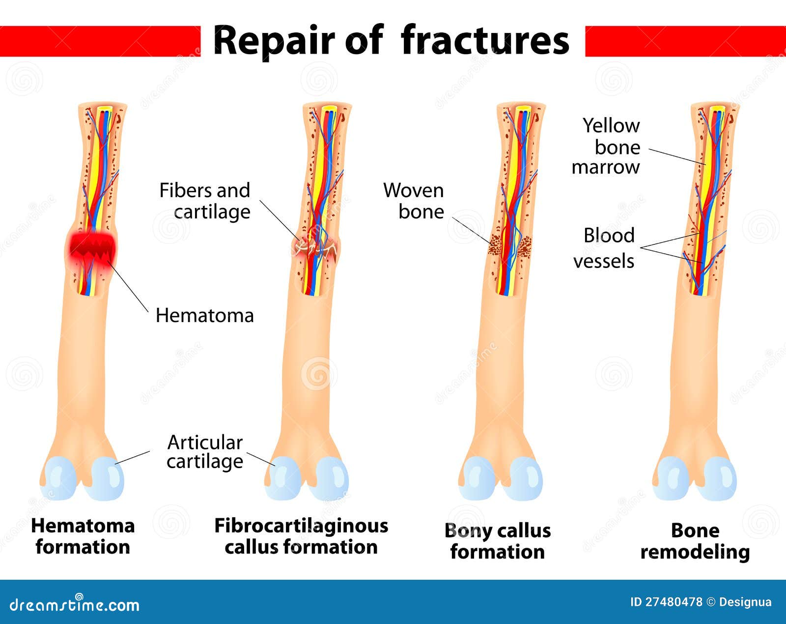
fracture healing steps

in march of 2015, american astronaut scottkelly and his russian colleague mikhail kornienko, began an unprecedented mission in space. they began a one-year term of service aboardthe international space station, the longest tour of duty ever served on the iss. now, i imagine there’s all sorts of stuffto worry about when you’re packing for a year-long space voyage, like, say, “howmany books should i bring? how many pairs of underwear? am i really okay with poopinginto a suctioned plastic bag every day for a year? will i come upon a derelict ship hauntedby some stranded and insane astronaut from a forgotten mission, like in pretty much everyspace horror movie ever? will there be coffee?â€
reasonable questions, all, but in reality,another one you might want to ask is: “will i be able to walk when i get back home?†we know micro-gravity is hard on a body, andthis mission is largely about testing the physical effects of being weightless for so long. astronauts often experience things like troublesleeping, puffy faces, and loss of muscle mass, but perhaps the most serious damage amicrogravity environment causes is to the bones. and bones, well, they’re pretty clutch. though they may look all dried up and austere,don’t be fooled -- your bones are alive. alive i tell you!
they’re actually as dynamic as any of yourorgans, and are made of active connective tissue that’s constantly breaking down, regenerating,and repairing itself throughout your lifetime. in fact, you basically get a whole new skeletonevery 7 to 10 years! in short, your bones do way more than justproviding your squishy sack of flesh with support and scaffolding and the ability tomove around. your bones are basically how you store thecalcium, phosphate, and other minerals you need to keep neurons firing and muscles contracting. they’re also crucial to hematopoiesis, orblood cell production. all of your new blood -- and we’re talking like, a trillion bloodcells a day! -- is generated in your bone
marrow, which also helps store energy as fat. bones even help maintain homeostasis by regulatingblood calcium levels and producing the hormone osteocalcin, which regulates bone formationand protects against glucose intolerance and diabetes. so, the big buzzkill about life in space isthat, up there, a person suffers one to two percent bone loss every month. by comparison, your average elderly personexperiences 1-2 percent bone loss every year. so for kelly and kornienko, that could meanlosing up to 20 percent over a year in orbit. given everything your bones do for you, that’sreally serious. and while most of that loss is reversibleonce they’re back on earth, it’s not as
easy as chugging some of madame pomfrey’sskel-e-gro potion. rehabilitation can take years of hard work,and that’s just after a few months in orbit… which is why kelly and kornienko are heroesof science, and not just for scholars of anatomy and physiology everywhere, but for anybodywho has bones. an average human body contains 206 bones,ranging in shape and size from the tiny stapes of the inner ear to the huge femur of thethigh. that’s a lot of bones to keep tabs on, soanatomists often divide these structures first by location, into either axial or appendiculargroups. as you might guess, your axial bones are foundalong your body’s vertical axis -- in your
skull, vertebral column, and rib cage. they’re kind of like your foundation, thestuff you can’t really live without -- they carry your other body parts, provide skeletalsupport, and organ protection. your appendicular bones are pretty much everythingelse, the bones that make up your limbs, and the things that attach those limbs to youraxial skeleton, like your pelvis and shoulder blades. these are the bones that help us movearound. from there, bones are generally classified by theirshape, and luckily those names are pretty obvious. long bones are your classic-looking, dog-bone-shapedbones -- the limb bones that are longer than they are wide, like tibia and fibula of your lower legs,but also the trio of bones that make up your fingers.
follow some of those long bones to your footor hand, and you’ll hit a cube-shaped short bone, like your foot’s talus and cuboid,or your wrist’s lacunate or scaphoid. your flat bones are the thinner ones, likeyour sternum and scapulae, and also the bones that make up your brain case. and your irregular bones are all the weirdly-shapedthings like your vertebrae and pelvis, which tend to be more specialized and unique. but despite their variations in size, shape,and finer function, all bones have a similar internal structure. they all have a dense, smooth-looking externallayer of compact, or cortical bone around
a porous, honeycomb-looking area of spongybone. this spongy bone tissue is made up of tinycross-hatching supports called trabeculae that help the bone resist stress. and it’salso where you typically find your bone marrow, which comes in two colors, red and yellow. red marrow is the stuff that makes blood cells,so you should be glad that you have some of that. and yellow marrow stores energy as fat -- ifyou happen to be a predatory animal, yellow bone marrow can be one of the best sourcesof calories you can find. the arrangement of these bone tissues, though, can beslightly different, from one type of bone to the next. in flat, short, and irregular bones, for example,these tissues kinda look like a spongy bone
sandwich on compact-bone bread. but in some of your classic long bones, likethe femur and humerus, the spongy bone and its red marrow are concentrated at the tips. these flared ends, or epiphyses bookend thebone’s shaft, or diaphysis, which -- instead of having spongy bone in the center -- surrounds a hollowmedullary cavity that’s full of that yellow marrow. now, although bone can look rock-solid, graba microscope and you’ll see that it’s actually loaded with layered plates and laced withlittle tunnels. it’s intricate and kinda confusing in there,but the more you zoom into the microanatomy of bones, the better you can see how they’re builtand how they function, right down to the cellular level.
let’s start with the basic structural unitsof bone, called osteons. these are cylindrical, weight-bearing structuresthat run parallel to the bone’s axis. look inside one and you’ll see that they’re composedof tubes inside of tubes, so that a cross-section of an osteon looks like the rings of a treetrunk. each one of these concentric tubes, or lamellae, isfilled with collagen fibers that run in the same direction but if you inspect the fibers of a neighboringlamella -- either on the inside or outside of the first one -- you’ll see that they run in adifferent direction, creating an alternating pattern. this reinforced structure helps your bone resisttorsion stress, which is like twisting of your bones, which they experience a lot, andi encourage you not to imagine what a torsion
fracture of one of your bones might feel like. now, bone needs nourishment like any othertissue, so running along the length of each osteon are central canals, which hold nervesand blood vessels. and then, tucked away between the layers oflamellae are tiny oblong spaces called lacunae. as tiny as they are, these little gaps arewhere the real work of your skeletal system gets done, because they house your osteocytes. these are mature bone cells that monitor andmaintain your bone matrix. they’re like the construction foremen of your bones, passingalong commands to your skeleton’s two main workhorses: the osteoblasts and the osteoclasts.
osteoblasts -- from the greek words for “boneâ€and “germ†or “sprout†-- are the bone-building cells, and they’re actuallywhat construct your bones in the first place. in the embryonic phase, bone tissue generallystarts off as cartilage, which provides a framework for your bones to grow on. whenosteoblasts come in, they secrete a glue-like cocktail of collagen, as well as enzymes that absorbcalcium, phosphate, and other minerals from the blood. these minerals form calcium phosphate, whichcrystallize on the cartilage framework, ultimately forming a bone matrix that’s about one-thirdmineral, two-thirds protein. from your time in the womb until you’reabout 25, your osteoblasts keep laying down more collagen and more calcium phosphate, untilyour bones are fully grown and completely hardened.
so while your osteoblasts are the bone-makers,your osteoclasts are the bone-breakers -- which is a kind of violent image. maybe think ofthem as like a bone-breaker-downer. although the two kinds of cells do exactopposite jobs, they’re not mortal enemies. in fact, i’m happy to report that they getalong fabulously, and create a perfect balance that allows your bones to regenerate. it’s like if you want to renovate your house,you’ve gotta rip out all those busted cabinets and the musty carpeting before you can bringin the nice hardwood floors and custom countertops. these cells work in a kinda similar way, ina process that i’d argue is less stressful than home improvement -- it’s called boneremodeling.
the supervisors of this process are thoseosteocytes, which kick things off when they sense stress and strain, or respond to mechanicalstimuli, like the weightlessness of space, or the impact of running on pavement. so, say you’re out running and somethinghappens -- nothing to be alarmed about! -- but suddenly the osteocytes in your femur detecta tiny, microscopic fracture, and initiate the remodeling process to fix it up. first, the osteocytes release chemical signalsthat direct osteoclasts to the site of the damage. when they get there, they secreteboth a collagen-digesting enzyme, and an acidic hydrogen-ion mixture that dissolves the calciumphosphate, releasing its components back into
the blood. this tear-down process is calledresorption. when the old bone tissue is cleaned out, theosteoclasts then undergo apoptosis, where they basically self-destruct before they can do anymore damage. but before they auto-terminate, they use the hormone hotline to call over the osteoblasts,who come in and begin rebuilding the bone. the ratio of active osteoclasts to osteoblastscan vary greatly, and if you stress your bones a lot, through injury, by carrying extra weight,or just normal exercise, those osteoclasts are going to be swinging their little wrecking ballsnon-stop, breaking down bone so it can be remade. in this way, exercising stimulates bone remodeling-- and ultimately bone strength -- so when you’re working out, you’re building boneas well as muscle.
which brings us back to our two space-heroes-slash-guinea-pigs, scott kelly and mikhail kornienko. space crews generally need to exercise atleast 15 hours a week to slow down the process of bone degradation, but even that can’tfully stave loss of bone density. in microgravity, osteocytes aren’t getting much loading stimuli, because less gravity means less weight. but, for reasons that we don’t understandyet, the osteoclasts actually increase their rate of bone resorption in low gravity, whilethe osteoblasts dial back on the bone formation. because there’s more bone breaking thanbone making going on, everything is out of balance, and suddenly people start experiencing1 to 2 percent monthly loss in bone mass. so, in addition to providing astronauts withoxygen and water and food and protection from
radiation and an environment that will keepthem mentally stable, it turns out that we also have to figure out how to keep theirbodies from consuming their own skeletons. but at least today we learned about the anatomyof the skeletal system, including the flat, short, and irregular bones, and their individualarrangements of compact and spongy bone. we also went over the microanatomy of bones,particularly the osteons and their inner lamella. and finally we got an introduction to theprocess of bone remodeling, which is carried out by crews of osteocytes, osteoblasts, andosteoclasts. special thanks to our headmaster of learningthomas frank for his support for crash course and free education. and thank you to all ofour patreon patrons who make crash course
possible through their monthly contributions.if you like crash course and you want to help us keep making cool new videos like thisone, you can check out patreon.com/crashcourse this episode was co-sponsored by the midnighthouse elves, fatima iqbal, and roger c. rocha crash course is filmed in the doctor cherylc. kinney crash course studio. this episode was written by kathleen yale, edited by blakede pastino, and our consultant, is dr. brandon jackson. our director is nicholas jenkins,the editor and script supervisor is nicole sweeney, our sound designer is michael aranda,and the graphics team is thought cafã©.
